Application of 3D Foot Scanners in Orthopedics
In the medical field, particularly in orthopedics, accurate diagnosis and treatment planning are key to ensuring patient recovery. With advancements in technology, various cutting-edge medical devices continue to emerge, providing doctors with more powerful tools to improve the quality of diagnosis and treatment. Among these, the 3D foot scanner, as an emerging technology, has shown tremendous potential in orthopedics. This article will explore how 3D foot scanners are transforming orthopedic practices and share specific cases of their clinical application.
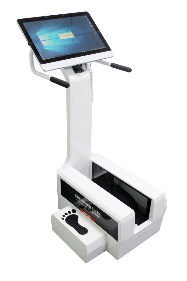
A 3D foot scanner is a technological device capable of quickly and accurately capturing the three-dimensional structural information of a patient’s feet. By using high-precision sensors and advanced image processing algorithms, the device generates a detailed digital model in a short period of time. This model includes not only basic measurements like foot length and width, but also more complex details such as arch height, toe alignment, and other structural features. These data are crucial for orthopedic doctors because they provide comprehensive insights into the patient’s foot condition, enabling the development of more precise treatment plans.
Firstly, 3D foot scanners play a vital role in the diagnostic phase. Many foot conditions such as flat feet, high arches, and bunions often require detailed examination to determine the underlying cause. Traditional methods may involve X-rays or CT scans, but these imaging techniques typically provide only two-dimensional images, which sometimes fail to fully reflect the actual condition. In contrast, the 3D foot scanner can generate a three-dimensional model, allowing doctors to examine the foot structure from multiple angles, making it easier to identify subtle problems. For example, in the evaluation of plantar fasciitis, the 3D scan can help doctors clearly see changes in the thickness of the plantar fascia and the condition of surrounding soft tissues, leading to a more accurate diagnosis.
Secondly, in surgical planning, 3D foot scanners are also indispensable. For cases that require complex reconstruction surgeries, such as congenital deformity correction or post-trauma repair, preoperative planning is critical. Using data obtained from the 3D scan, doctors can simulate different surgical approaches on a computer and predict the outcome of each option. This not only helps in selecting the best treatment method but also reduces surgical risks and increases success rates. Additionally, based on these detailed data, personalized implants or orthotics can be custom-designed for better therapeutic outcomes.
Beyond surgical treatment, 3D foot scanners also play an important role in non-surgical therapies. For example, in the production of orthotic insoles, traditional manual measurement methods are often imprecise, leading to insoles that may not meet the patient’s specific needs. With 3D scanning technology, insoles can be designed based on the unique foot shape of each patient, effectively alleviating pain and improving gait. This is particularly important for patients with chronic foot conditions, as wearing ill-fitting insoles can worsen their symptoms over time.
It is worth noting that the application of 3D foot scanners is not limited to adult patients. Children and adolescents, who are still in the process of growth and development, experience rapid changes in foot structure, making regular monitoring essential. By conducting regular 3D foot scans, doctors can detect potential problems early and intervene before more serious complications arise. Additionally, this technology can be used for research purposes to help scientists better understand foot development patterns in different age groups, providing a scientific basis for prevention and treatment.
Despite the many advantages of 3D foot scanners, some challenges remain in their practical application. First is the cost issue. Purchasing and maintaining such high-tech equipment requires a significant financial investment, which may limit its widespread use in certain medical institutions. Second is the issue of data security and privacy protection. Since the information involves personal biometric data, strict measures must be taken to ensure the secure storage and transmission of the data. Lastly, specialized training for healthcare professionals is necessary to ensure they can operate the equipment proficiently and interpret the scan results accurately.
The 3D foot scanner has become an indispensable tool in orthopedics. It not only enhances diagnostic accuracy but also optimizes the design and execution of treatment plans. Whether from the perspective of improving patients’ quality of life or advancing medical research, this technology is of great significance. As related technologies continue to improve and costs gradually decrease, we have reason to believe that 3D foot scanners will be widely adopted by more medical institutions, bringing benefits to more patients.
The application of 3D foot scanners in orthopedics marks a significant advancement in medical technology. It not only improves diagnostic precision but also offers new possibilities for personalized treatment. Through this advanced technology, doctors can better understand the condition of patients’ feet and create more effective treatment plans. As the technology continues to develop, we hope that 3D foot scanners will benefit more patients and positively impact their health and quality of life.
