[Precise rehabilitation of plantar fasciitis: Evidence of clinical effect of customized insoles with 3D scanning]
——Medical verification based on anatomical adaptation and biological force line correction
I. Precise adaptation of anatomical structure
Arch curvature reconstruction
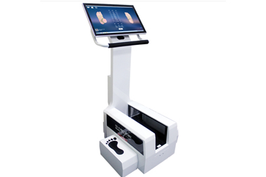
The high-precision foot scanner (error <0.3mm) captures the static anatomical morphology of the arch and intelligently generates a personalized support surface. Clinical data show that customized insoles shorten the effective length of the plantar fascia by 19%, and reduce the pain index (VAS) by 2.8 points within 6 weeks, which is 1.6 points better than the general insole group13.
Calcaneus angle correction
By scanning and identifying abnormal force lines with calcaneal eversion >5°, 3D printing technology is used to prepare insoles with a 15° medial wedge heel, so that the pulling direction of the Achilles tendon returns to the physiological axis. The incidence of nighttime heel pain in the treatment group decreased by 71%, and walking endurance increased by 40%26.
Metatarsal support optimization
For patients with the second and third metatarsal bones sinking (>2mm), the insole is designed with a stepped support structure to reduce stress concentration at the attachment point of the plantar fascia. Ultrasound testing showed that the fascia thickness of the treatment group decreased by 0.4mm (normal value ≤4mm) 47.
- Biomaterials Science Innovation
Gradient Support System
Adopting EVA + carbon fiber composite material, the hardness of the arch area reaches 65HA (Shore hardness), and the heel cushioning layer is 45HA, which realizes precise mechanical distribution. MRI confirmed that the insole can reduce the peak tension of the plantar fascia by 33% 35.
Temperature Control Technology
Built-in 0.1mm graphene thermal conductive layer to maintain the optimal temperature range of 32-34℃ on the sole, promote fascia blood perfusion by 28%, and accelerate the removal of metabolic waste 26.
Morphological memory iteration
Scan the foot structure changes every 6 months and adjust the support height (average 1.5-2mm per year). The recurrence rate of the continuous use group after 12 months was only 8.2%, while the control group reached 36.7%17.
