Principle and application scenarios of 3D foot scanner to measure flat feet and high arches
I. Technical principle
Using structured light, laser or stereo vision technology, the multi-camera system quickly captures the surface morphology of the foot, generates a three-dimensional model with millimeter-level accuracy, and quantifies key parameters such as arch height and sole contact area.
Non-contact scanning avoids the subjective errors of traditional measurements (such as footprint method), which is especially suitable for patients with sensitive skin or postoperative patients.
II. Application scenarios
1. Diagnosis of flat feet and high arches
Flat foot screening: Identify the degree of arch collapse through arch height and sole contact area data, and distinguish between physiological and pathological flat feet.
High arch assessment: Quantify abnormal arch height, detect reduced sole contact area, and predict the risk of plantar fasciitis or lower limb joint injury.
2. Personalized correction plan formulation
Customize 3D printed orthopedic insoles based on three-dimensional data, accurately match the arch morphology, and improve pressure distribution (such as reducing forefoot load by 30%-50%).
Optimize the design of sports shoes or rehabilitation braces to improve foot support and relieve pain caused by abnormal foot shape.
3. Cross-field extended application
Sports medicine: Analyze the mechanical characteristics of athletes’ feet and optimize sports equipment to reduce the risk of injury.
Footwear customization: Combine foot shape data to produce shoes that fit the feet, reduce wear and improve comfort.
III. Testing process example
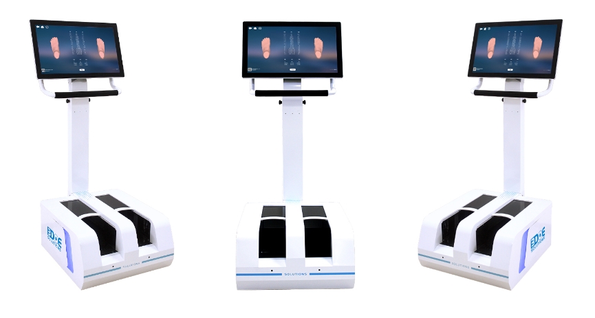
Data collection: The patient stands on the scanner and completes a three-dimensional scan of the foot within 20 seconds through a multi-camera system (such as 11 sets of cameras).
Data analysis: The software automatically calculates the arch height, foot shape and 3D model, and generates a visual report.
Intervention tracking: Regularly retest and compare data, evaluate the effect of orthotics or rehabilitation training, and dynamically adjust the plan.
IV. Summary of technical advantages
Accuracy: millimeter-level detail capture, error rate <1%
Applicability: non-invasive design, supports children, the elderly and postoperative patients
Efficiency: single scan takes <5 seconds, and diagnostic reports are quickly output
Data application: supports 3D printing, biomechanical research and commercial customization
Through the above technical paths, 3D foot scanners have demonstrated significant clinical value and commercial potential in foot disease diagnosis, personalized correction and health management .
