In spinal health management, adolescent scoliosis is receiving increasing attention from both families and schools. When parents take their children for an examination, the two most commonly recommended methods are: X-ray imaging and 3D spinal scanner testing.
So here comes the question — both are tools for examining spinal issues, but which is safer? Is there a big difference in effectiveness? Can they replace each other?
Today, let’s explain everything clearly at once!
1. First, understand the main characters: X-ray and 3D scanner
X-ray examination is currently one of the most authoritative methods used in hospitals to diagnose scoliosis. It uses ionizing radiation to penetrate the body and clearly image the alignment of the spine, allowing precise measurement of the curvature angle (known as the Cobb angle). It is commonly used for initial diagnosis, preoperative evaluation, and follow-up during treatment.
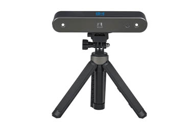
The 3D spinal scanner, on the other hand, is a non-invasive, radiation-free surface scanning system. Using laser or infrared technology, it performs a holographic scan of the back, reconstructing the surface contour and spatial posture of the spine. It then evaluates the tendency of scoliosis and the symmetry of areas like the shoulder blades and pelvis. It is suitable for screening, monitoring, and dynamic observation.
2. Comparison of safety: 3D scanner wins completely
In terms of safety, the 3D spinal scanner is clearly superior, for two reasons:
1. No radiation
The 3D scanner does not use X-rays and thus poses no risk of ionizing radiation. It is especially suitable for children and adolescents, and even with multiple scans per year, there are no health concerns.
2. No contact, non-invasive
The entire scanning process requires no undressing, no adhesive patches, and causes no discomfort. For children who are afraid of hospitals, the experience is gentler and more friendly.
In comparison, although X-rays use low-dose radiation, if taken frequently — especially during the growth period of children — the accumulated radiation may pose potential risks. Therefore, international guidelines typically recommend limiting the frequency of X-rays and using them only at key diagnostic points.
3. Comparison of effectiveness: complementary functions, different focuses
X-rays can “see the bones,” with the advantage of clear imaging and precise measurement of skeletal structures. However, they provide only 2D images and cannot show muscle balance or body posture dynamics.
The 3D scanner “sees the posture,” showing clearly whether the shoulders are level, whether the pelvis is tilted, and whether the spinal surface has rotated. It is a good tool for dynamic posture assessment but cannot view internal bone details, and therefore cannot replace X-rays for definitive diagnosis.
So, the two are not in a competitive relationship, but rather complement each other and work together in clinical diagnosis and treatment.
