Regarding the instruments used to assess flat feet, the methods commonly used in clinical and rehabilitation departments have become increasingly digitalized and precise. Among them, the Foot 3D Scanner has become an increasingly common assessment tool. Below is a classification of instruments used to assess flat feet, with a focus on the Foot 3D Scanner:
I. Common Instruments and Methods for Assessing Flat Feet
1. Foot Pressure Plate
Function: Measures plantar pressure distribution during standing or walking.
Application: Determines the degree of arch collapse and whether pressure is biased toward the medial or lateral side.
Advantages: Allows dynamic gait analysis; suitable for runners, postoperative evaluation, etc.
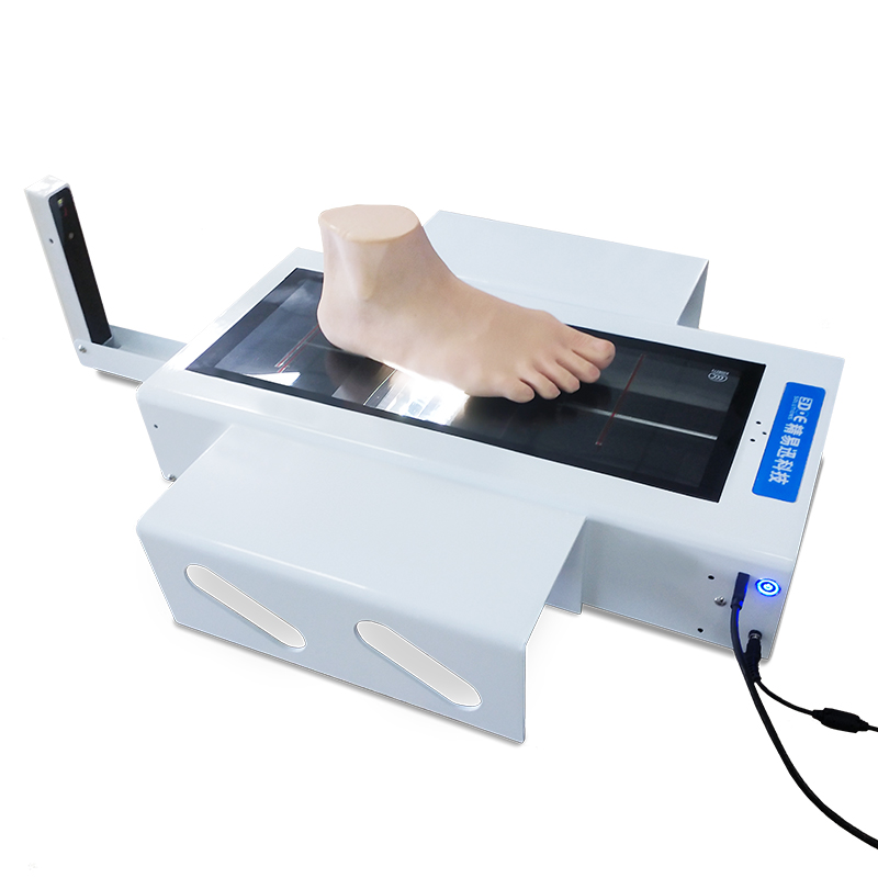
2. Foot 3D Scanner
Function: Captures three-dimensional structural data of the plantar and dorsal aspects of the foot to reconstruct the arch shape.
Application: Accurately assesses arch height and foot structure, used to diagnose flat feet, high arches, overpronation, etc.
Advantages:
Non-invasive, rapid imaging
Suitable for custom orthotic insoles or orthopedic shoes
Foot data can be stored for follow-up and comparison
Applicable population: Children, adolescents, patients with foot disorders, athletic individuals, etc.
3. Static Footprint Device
Function: Leaves a plantar imprint by standing on a pressure board to preliminarily assess the arch condition.
Application: Widely used in school screenings or primary care assessments.
Advantages: Low cost, easy to operate
Disadvantages: Only suitable for static evaluation, limited precision
4. X-ray (Standing Lateral View of the Foot)
Function: Assesses medial arch height and talonavicular alignment from a bony structure perspective.
Advantages: Applicable for moderate to severe flat feet, especially for preoperative evaluation or skeletal deformity screening.
Note: Involves radiation exposure, used only when medically necessary
II. Working Principle and Advantages of the Foot 3D Scanner
Principle
The Foot 3D Scanner uses laser or structured light to quickly scan the foot and generate a high-precision 3D digital model. It extracts data such as arch height, foot length, foot width, interphalangeal angles, and heel deviation.
Advantages
No skin contact required, completes scanning in a few seconds
Accurately reconstructs foot contour with minimal error (typically less than 1 mm)
Visualized reports help doctors and patients better understand the foot condition
Can be used to assist in the fabrication of personalized orthotic insoles
III. Common Process of Assessing Flat Feet (Using Instruments)
Initial consultation and visual foot assessment by physician or rehabilitation therapist
Static plantar pressure test (to observe midfoot collapse)
3D foot scan to obtain arch height and deviation angles
X-ray examination if necessary, to confirm skeletal structure
Provide diagnostic conclusion and recommend whether intervention (such as orthotic insoles or rehabilitation training) is needed
