I. Equipment Coordination Workflow
- 3D Scanning Phase
Optical scanners capture static structural data:
High-precision reconstruction of arch height, foot length/width ratios, and toe alignment parameters
Identification of deformities (e.g., hallux valgus >15° or abnormal high-arch indices)
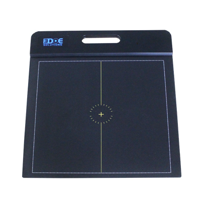
- Pressure Testing Phase
Pressure plates detect dynamic functional performance:
Analysis of plantar pressure distribution during gait cycles (COP trajectory deviation >5mm indicates abnormalities)
Calculation of Arch Index (AI >0.21 suggests flat foot risk)
II. Key Integrated Evaluation Indicators
- Structure-Function Correlation Analysis
Cross-validate arch height from scans with AI from pressure tests:
High arch in structure but normal AI → suggests compensatory functional adaptation
Normal structure but abnormal AI → requires examination of muscle imbalances
- Orthotic Solution Validation
Post-custom orthotic fabrication based on 3D scans, efficacy tested via pressure plate:
≥20% reduction in midfoot pressure deemed effective
Gait symmetry improvement (stride difference <10%)
III. Clinical Application Scenarios
- Sports Rehabilitation
Combine bone structure changes from scans with gait anomalies from pressure plates to identify injury mechanisms in athletes - Pediatric Foot Development
Track scan data and pressure parameters every 6 months to establish arch development curves (excluding growing pains) - Diabetic Foot Screening
Use pressure plates to detect high-pressure zones (>300kPa), then precisely locate ulcer risk points with 3D scans
The integrated solution improves detection accuracy by 35% compared to single-device methods, particularly for complex foot conditions requiring customized interventions.
