In the fields of modern rehabilitation medicine and personalized health management, orthopedic insoles serve as important assistive devices for improving gait abnormalities, relieving foot pain, and preventing lower limb injuries. Their effectiveness highly depends on how well they match the user’s foot morphology.
Traditional methods such as manual casting or two-dimensional measurements suffer from limitations including large errors, time-consuming processes, and difficulty in reproducing complex curved surfaces, making them increasingly unable to meet the growing demand for precision healthcare.
With the advancement of digital technology, foot shape 3D scanners are gradually becoming core tools in orthopedic insole design, driving orthopedic aids from an era of “experience-based customization” into a new era of “data-driven precise fitting.”
- Limitations of Traditional Methods and the Necessity of Digital Transformation
In the past, manufacturing orthopedic insoles largely relied on plaster casting or foam impression methods. Although these methods could roughly reflect the foot contour, they had many drawbacks: plaster is prone to deformation during solidification, and demolding may cause structural distortion; foam boards experience uneven pressure, failing to accurately represent plantar pressure distribution.
Moreover, the process is cumbersome and provides a poor user experience, especially unfriendly to children, elderly individuals, or postoperative patients. More importantly, these analog data must be manually converted into digital models, involving multiple intermediate steps that lead to significant loss of accuracy, ultimately resulting in poor insole fit and reduced corrective effectiveness.
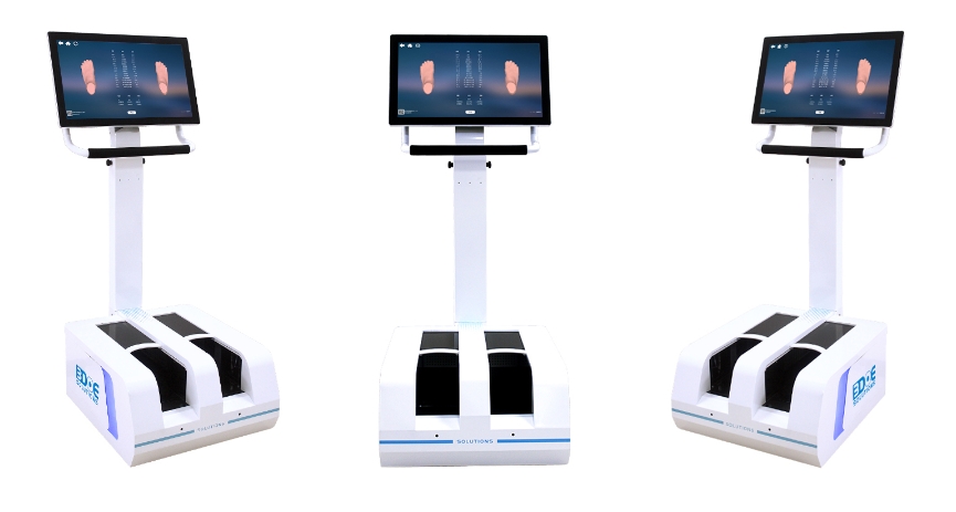
In contrast, foot shape 3D scanners use non-contact optical technology to acquire tens of thousands of spatial coordinate points on the foot surface within seconds, generating high-precision 3D digital models.
This process is not only fast and comfortable but also fully captures key anatomical features such as arch curvature, heel tilt angle, forefoot width, and toe bone alignment, providing a reliable data foundation for subsequent personalized design.
- How 3D Scanning Empowers Precise Orthopedic Insole Design
High-Fidelity Data Acquisition
Modern foot scanners typically use structured light or stereo vision technology to scan the foot under static standing (weight-bearing) conditions. The foot shape under weight-bearing best reflects biomechanical characteristics during actual walking, especially the degree of arch collapse and plantar pressure distribution. The 3D model can precisely quantify parameters such as arch height, varus/valgus angles, and longitudinal arch index, providing objective criteria for diagnosing conditions like flat feet, high arches, and intoeing.
Intelligent Analysis and Automatic Annotation
Companion software can utilize artificial intelligence algorithms to automatically identify key anatomical landmarks on the foot, such as the navicular tuberosity, center of the calcaneus, and the first and fifth metatarsal heads. Based on these reference points, the system calculates inclination angles, rotational status, and alignment relationships across different foot regions, assisting rehabilitation physicians or orthotists in making professional diagnoses.
Personalized Insole Modeling
Based on the 3D foot model, designers can perform virtual modifications according to the patient’s specific needs. For example, adding medial longitudinal arch support for patients with flat feet, designing higher and stiffer heel cups for those with hindfoot valgus, or reserving pressure-relief zones for diabetic feet. All modifications can be previewed in real time within the software, ensuring structural rationality and smooth boundaries.
Seamless Integration with Digital Manufacturing
After design completion, the data can be directly imported into CNC machines or 3D printers for fabrication, enabling full digitalization of the entire “scanning–design–production” process. This integrated workflow significantly shortens delivery cycles while ensuring each insole strictly corresponds to the original data, eliminating human error.
- Clinical Value and Application Prospects
In rehabilitation centers, foot and ankle specialty clinics, and sports medicine institutions, foot shape 3D scanning has become the standard preliminary step in custom orthopedic insole production. It not only enhances correction effectiveness but also improves patient compliance—when users see their own foot models and customized solutions, they better understand the treatment principles and develop greater trust.
Additionally, 3D data can be archived long-term, facilitating tracking of foot morphology changes, evaluating intervention outcomes, and providing continuous support for chronic disease management (such as diabetic foot and rheumatoid arthritis).
