The application of a three-dimensional foot laser scanner to arch detection is a representation of the deep integration of modern biomechanics and digital measurement technology. Its main function is to achieve high-precision detection of foot structure and conduct comprehensive quantitative analysis, providing scientific evidence for clinical diagnosis, orthotic design, and sports rehabilitation.
The foot arch is an important supporting structure of the human foot. The shape and function of the arch directly affect the transmission of lower limb mechanics, gait stability, and the risk of sports injuries. Therefore, accurately evaluating the arch is of great significance in both medicine and kinesiology.
Traditional methods of arch detection mostly rely on footprint measurement or X-ray imaging analysis, which have problems such as limited accuracy, simple information, or being affected by human operation.
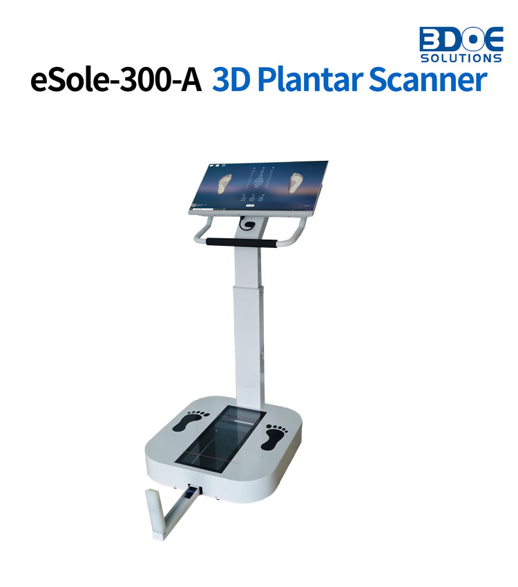
In contrast, the three-dimensional foot laser scanner adopts a non-contact scanning method, which can collect panoramic data of the foot surface in a short time, and accurately reconstruct the foot contour in the form of a three-dimensional point cloud.
This technology can simultaneously obtain multidimensional parameters such as arch height, arch length, arch width, and arch inclination angle, providing quantification for the judgment of arch types, structural integrity, and potential abnormalities.
Specifically, the three-dimensional foot laser scanner has the following functions in arch detection:
First, strong accuracy.
Laser scanning technology can achieve sub-millimeter resolution and has sensitivity to changes in foot surface concavity and convexity, arch curvature, and other microstructures, thereby ensuring the objectivity and reliability of the measurement results.
Second, high repeatability.
The scanning process is highly automated, reducing human operation errors, making continuous measurements or long-term follow-up data highly comparable, thus laying a foundation for dynamic clinical observation.
Third, data visualization.
Through the reconstruction of a three-dimensional model, the arch shape can be displayed intuitively. Doctors or researchers can evaluate the foot structure from multiple angles and assist in the development of personalized orthotic insoles or rehabilitation training programs.
In the end, it facilitates digital analysis and intelligent processing.
Three-dimensional foot scanning data can be input into computer algorithms for mechanical simulation, gait analysis, or prediction of foot pressure distribution, thereby providing scientific evidence for the prevention and treatment of foot diseases, the evaluation of sports injuries, and rehabilitation interventions.
